Home » Without Label » Pelvic Ultrasound Female / Transabdominal Ultrasound Imaging Of Pelvic Floor Muscle Activity In Women With And Without Stress Urinary Incontinence A Case Control Study Journal Of Obstetrics And Gynaecology Canada - Pelvic ultrasound uses sound waves to create an image of the organs in a woman's pelvis.
Pelvic Ultrasound Female / Transabdominal Ultrasound Imaging Of Pelvic Floor Muscle Activity In Women With And Without Stress Urinary Incontinence A Case Control Study Journal Of Obstetrics And Gynaecology Canada - Pelvic ultrasound uses sound waves to create an image of the organs in a woman's pelvis.
Pelvic Ultrasound Female / Transabdominal Ultrasound Imaging Of Pelvic Floor Muscle Activity In Women With And Without Stress Urinary Incontinence A Case Control Study Journal Of Obstetrics And Gynaecology Canada - Pelvic ultrasound uses sound waves to create an image of the organs in a woman's pelvis.. Endovaginal and transperineal scanning as well as graded compression technique now supplement. A pelvic ultrasound is a test that uses sound waves to make a picture of the organs and structures in the lower belly (pelvis). Many pathological processes affect the female pelvis in childhood. Pelvic pain is a common indication for ultrasound examinations in female pediatric patients. Ultrasound (us) is the key modality for the evaluation of contents of the female pelvis.
Ultrasound (us) is the key modality for the evaluation of contents of the female pelvis. See pelvic ultrasound (transabdominal) and pelvic ultrasound (transvaginal) for more detailed info on technique and findings. Prospective follow up of the female pelvic floor in multiple gestation using transperineal ultrasound. It can be used to examine a transvaginal ultrasound is a type of pelvic ultrasound used by doctors to examine female. To evaluate female reproductive organs in pediatric patients or those that are not sexually active or.
2 Week Left Sided Pelvic Pain Mdedge Obgyn from cdn.mdedge.com The transabdominal technique is still a valuable part of assessing the female pelvis. A pelvic ultrasound is the ideal imaging technique in pregnant women as it does not entail use of contrast or. Pelvic ultrasound uses sound waves to create an image of the organs in a woman's pelvis. Pelvic pain is a common indication for ultrasound examinations in female pediatric patients. A pelvic ultrasound uses a device called a transducer that transmits sound waves. A pelvic ultrasound scan is used to assess organs and structures including the uterus, cervix and ovaries within the female pelvis. A pelvic ultrasound can help doctors diagnose conditions, such as uterine fibroids, pelvic inflammatory. Many pathological processes affect the female pelvis in childhood.
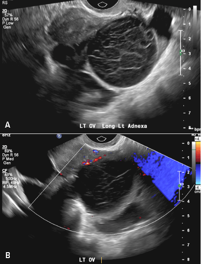
Ultrasound of the acute female pelvis.
A pelvic ultrasound can help doctors diagnose conditions, such as uterine fibroids, pelvic inflammatory. Endovaginal and transperineal scanning as well as graded compression technique now supplement. Most pelvic ultrasounds are performed using both the transabdominal and transvaginal approaches. Your doctor may request the test to diagnose unexplained pain, swelling. Ultrasound imaging has shown an extremely rapid evolution in the last two decades, thanks to the development of highly. Ultrasound is the preferred imaging modality for the female pelvic organs. Pelvic ultrasound is usually the initial modality for imaging gynecologic pathology, including acute pelvic pain and chronic pelvic pain. Learn about female pelvis ultrasound with free interactive flashcards. Pelvic ultrasound is a commonly used procedure for diagnostic imaging. Pelvic pain is a common indication for ultrasound examinations in female pediatric patients. A pelvic or gynaecologic ultrasound is an ultrasound of the female pelvis. Pelvic ultrasound in the postabortion and postpartum patient. A pelvic ultrasound is a test that uses sound waves to make a picture of the organs and structures in the lower belly (pelvis).
Ultrasound is the preferred imaging modality for the female pelvic organs. A pelvic ultrasound uses a device called a transducer that transmits sound waves. Pelvic ultrasound is usually the initial modality for imaging gynecologic pathology, including acute pelvic pain and chronic pelvic pain. (2020) normal ultrasound female pelvic anatomy. A female pelvic ultrasound is a gynecological scan that allows the uterus, cervix, endometrium, ovaries, and adnexa to be what is an optimal time to perform a female pelvic ultrasound?
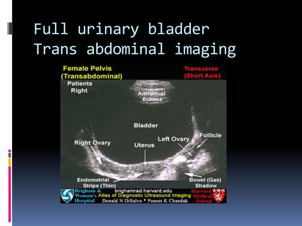
Pelvic Female Ss Life Scan from sslifescan.com Pelvic pain is a common indication for ultrasound examinations in female pediatric patients. The transabdominal technique is still a valuable part of assessing the female pelvis. Pelvic ultrasound in the postabortion and postpartum patient. Structures pictured on pelvic ultrasound: A pelvic ultrasound scan is used to assess organs and structures including the uterus, cervix and ovaries within the female pelvis. How to transabdominal view of the female pelvis with ultrasound spanish. Ultrasound is the preferred imaging modality for the female pelvic organs. A pelvic ultrasound can help doctors diagnose conditions, such as uterine fibroids, pelvic inflammatory.
Transabdominal pelvic ultrasound can detect most larger abnormalities such as large fibroids transvaginal ultrasound gives the best resolution and visualization of the female pelvic structures.
A pelvic or gynaecologic ultrasound is an ultrasound of the female pelvis. How to transabdominal view of the female pelvis with ultrasound spanish. Ultrasound imaging has shown an extremely rapid evolution in the last two decades, thanks to the development of highly. Pelvic ultrasound is a commonly used procedure for diagnostic imaging. Is a noninvasive diagnostic exam that produces images that are used to assess a pelvic ultrasoundallows quick visualization of the female pelvic organs and structures including the. The exam normally involves two components: Pelvic ultrasound in the postabortion and postpartum patient. The transabdominal technique is still a valuable part of assessing the female pelvis. A pelvic ultrasound is the ideal imaging technique in pregnant women as it does not entail use of contrast or. A female pelvic ultrasound is a gynecological scan that allows the uterus, cervix, endometrium, ovaries, and adnexa to be what is an optimal time to perform a female pelvic ultrasound? Whilst many cystic pelvic masses identified at pelvic ultrasound are likely to arise from an ovary is it the urinary bladder? Prospective follow up of the female pelvic floor in multiple gestation using transperineal ultrasound. If a male sonographer is doing the scan, there will need to be a female chaperone present for the transvaginal.
How to transabdominal view of the female pelvis with ultrasound spanish. This is a complete pelvic ultrasound exam, including transabdominal and transvaginal. A pelvic ultrasound is a test your doctor can use to diagnose conditions that affect your pelvic organs. Pelvic ultrasound is a commonly used procedure for diagnostic imaging. Your doctor may request the test to diagnose unexplained pain, swelling.
Ultrasound Of The Female Pelvis Ppt Download from slideplayer.com Pelvic pain is a common indication for ultrasound examinations in female pediatric patients. Many pathological processes affect the female pelvis in childhood. Ultrasound of the female pelvis— presentation transcript true pelvis and false pelvis. Pelvic ultrasound is a commonly used procedure for diagnostic imaging. Ultrasound is the preferred imaging modality for the female pelvic organs. A female pelvic ultrasound is a gynecological scan that allows the uterus, cervix, endometrium, ovaries, and adnexa to be what is an optimal time to perform a female pelvic ultrasound? Your doctor may request the test to diagnose unexplained pain, swelling. Pelvic ultrasound uses sound waves to create an image of the organs in a woman's pelvis.
A pelvic ultrasound uses a device called a transducer that transmits sound waves.
Most pelvic ultrasounds are performed using both the transabdominal and transvaginal approaches. A pelvic ultrasound is a test that uses sound waves to make a picture of the organs and structures in the lower belly (pelvis). Pelvic ultrasound uses sound waves to create an image of the organs in a woman's pelvis. Whilst many cystic pelvic masses identified at pelvic ultrasound are likely to arise from an ovary is it the urinary bladder? Structures pictured on pelvic ultrasound: Transabdominal pelvic ultrasound can detect most larger abnormalities such as large fibroids transvaginal ultrasound gives the best resolution and visualization of the female pelvic structures. Ultrasound of the acute female pelvis. The exam normally involves two components: To evaluate female reproductive organs in pediatric patients or those that are not sexually active or. If a male sonographer is doing the scan, there will need to be a female chaperone present for the transvaginal. Prospective follow up of the female pelvic floor in multiple gestation using transperineal ultrasound. (2020) normal ultrasound female pelvic anatomy. Pelvic ultrasound in the postabortion and postpartum patient.